Purification of total cell DNA: 7th april

Purification of DNA

DNA is the basic constituent of any living being, and some viruses too. The isolation and purification of DNA is a significant step in any molecular biological technique. We are to discuss about the mentioned three.

  • Total cell DNA preparation
  • Preparation of plasmid DNA
  • Preparation of bacteriophage DNA

Preparation of Total Cell DNA

The procedure can be divided into the following 4 steps

Step 1: Bacterial culture growth and harvest

Step 2: Cell wall rupture and preparation of cell extract

Step 3: Purification of DNA from the cell extract

Step 4: Concentration of DNA solution

Bacterial culture and growth

Most of the bacteria are grown in or are preferred to grow in liquid media {broth culture}.

  • The culture medium must provide 2 balanced mixtures of essential nutrients at concentrations that will allow the bacteria to grow and divide effectively
  • Though the medium are of different types, we are here to discuss only about nutrient media.
  • The media should consider the basic sources
  • The most frequently used are
      • Defined media

In this case we know the exact concentration of all the chemicals used in the media preparation. This media contains 2 mixtures of inorganic nutrients to provide essential elements like N, Mg, Ca as well as glucose. Additional growth factors like trace elements and vitamin may be added to provide additional nutrients to the bacteria depending upon the bacterial species.

Example

Composition of M9 medium

Constituent

Amount

Source for?

NaH2PO4

6

Sodium source

KH2PO4

3

Potassium source

Nacl

0.5

Salt

NH4Cl

1.0

MgSO4

0.5

Glucose

2.0

CaCl2

0.015

Undefined Media

In this case the precised identity and the quantity of its components are not known. Ingredients like tryptone and yeast extract are used which are complicated mixture of unknown chemical compounds. It is known as tryptone contains small peptides and amino acids like whereas yeast extract supplement provides N2 and glucose requirement to the bacteria. This complex media needs no further supplementation and support growth of wide range of bacterial species

Constituent

Amount req

Source

Tryptone

10

Yeast extract

5

Nacl

10

Preparation of cell extract

The bacterial cell is enclosed ni a cytoplasmic membrane and surrounded by 2 rigid cell wall. In some species like E.Coli, the cell wall itself may be engulfed by the second outer membrane. All these barriers have to be disrupted to release the cell components which can be done by 2 methods.

  • Physical method (Use of beads and other physical methods to rupture the cell wall)
  • Chemical method
    • Generally chemical method is used by making use of EDTA or lysozyme (secreted by saliva).
    • Lysozyme is present in secretion by saliva, tears egg-white which digests the polymeric compounds giving rigidity to the cell wall.
  • EDTA (Ethylene Diamine Tetra-acetic Acid), it removes the Mg2+ ions that are essential for preserving the overall structure of the cell.

It also inhibits the cellular enzymes that could degrade the DNA.

  • SDS (Sodium Dodecyl Sulphate).
    • In some cases it is also added, which helps in the process of lysis by removing lipid molecules. Generally mixture of EDTA and lysozyme is used.
    • This mixture is then centrifuged at a speed of 8000 rpm for 110 mins. It causes the precipitation of cell forming and the pellets in the test-tube.
    • Finally the cell extracts are extracted from cell debris, which are pellet down by centrifugation

Purification of DNA from cell extract

  • The standard way to deproteinize a cell is to add phenol or a 1:1 mixture of phenol and chloroform.
  • The organic solvents precipitate proteins but leave the nucleic acids (DNA and RNA) in an aqueous solution.
  • The result is that is the cell extract is mixed gently with the solvent, and the layers then separated by centrifugation, precipitated protein molecules are left as a white coagulated mass at the interface between the aqueous and organic layers.
  • The aqueous solution of nucleic acids can then be removed with a white pipette.
  • Cell extract is treated with protease such as pronase or proteinase K before extraction.
  • These enzymes will break polypeptides into smaller units thus making phenol easier to remove them.
  • The only effective way to get rid of RNA is the use of Ribonuclease enzyme which will rapidly degrade thee molecules into ribonucleotide subunits.

Concentration of DNA samples

  • The most frequently used method of concentration is ethanol precipitation.
  • In the presence of salt and a temperature of -20c or less absolute ethanol with efficiently precipitate polymeric nucleic acids.
  • With 2 thick solution of DNA, the ethanol can be layered on the top of the sample.
  • A spectacular trick is to push a glass rod through the ethanol into the DNA solution.
  • When the rod is removed, DNA molecules will adhere and be pulled out of the solution in the form of long fiber.
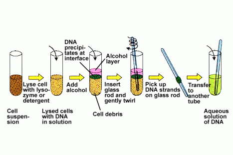
DNA out of a glass rod

for further reading click HERE

Vector: Bacteriophage lambda and M13: 7th april

Bacteriophage

Definition

They are one of the types of virus that attacks the bacteria and infects it.

It was first reported by Frederick Twort, a british biologist and later by Felix d’Herelle, a French microbiologist.

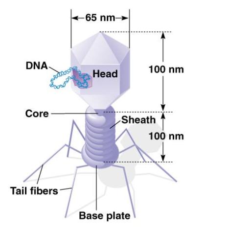
Diagram of phage

phage 1

Characteristic features of a bacteriophage

  1. They have a typical structure of outer protein capsid enclosing a genetic material
  2. The genetic material can be DNA or RNA and might be ss or ds.
  3. The size of the nucleotides can vary from 5000 to 500, 000 nucleotides
  4. The genetic material can be circular or linear
  5. They are much smaller than the bacteria typically of size 20nm to 200nm.
  6. The prime source of these virions is the highly populated areas of bacteria, mostly the sea water where upto 9*108 virion per ml is found.

The classification

They are classified by the International Committee on Taxonomy of Viruses (ICTV) according to the morphology and characteristics.

Replication

There are two stages of bacteriophage replication

  1. lytic cycle
  2. lysogenic cycle

Lytic cycle

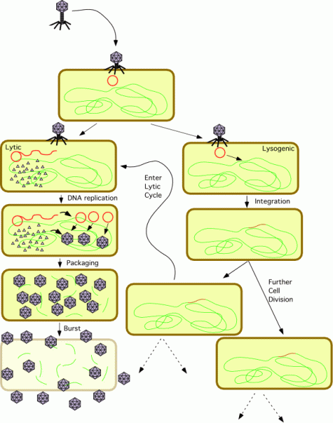
In this case the phage particle infects the bacterial cell and inserts the viral genetic material into it. The viral genetic material if is RNA uses reverse transcriptase enzyme to convert it to DNA and thus translates other building materials for a potential virion. The number of this virions increase at an exponential rate to burst the bacterial cell and set free to attack other baterial cells in the vicinity. This is a pretty fast rate of replication.

Lysogenic cycle

In this case the viral genetic material is incorporated into the bacteria’s own chromosome and thus is transferred to its progeny. When integrated with the host DNA the formation is called a prophage. And the phages able to cause lysogeny are called temperate phages. The virus remains dormant until the host conition deteriorates and once the stage reaches the viral DNA becomes active and thus causes lysis of the cell.

The prophages sometimes add more functions to the host cell. This is called lysogenic conversion. One famous example is the conversion of harmless viobrio cholerae by a phage into a highly virulent one. This is one reason why temperate phages are not used for phage therapy.

lytic and lysogenic

Diagram explaining the lytic and lysogenic cycle of a bacteriophage

More information at

click HERE


One more classification of bacteriophage is a m13 bacteriophage

M13 Bacteriophage

  1. It is a filamentous bacteriophage composed of circular single stranded DNA
  2. The length is 6407 nucleotides
  3. It is encapsulated in 2700 copies of the major coat protein P8, capped with 5 copies of two different minor coat proteins (P9, P6, and P3).
  4. The minor P3 attaches to the receptor at the tip of the host E.Coli.
  5. It is not lethal but causes plaques in the bacterial cell
  6. It is a non-lytic virus
  7. The M13 phage is used for many recombinant DNA processes due to its extreme size and the virus has also been studied for its uses in nanostructures and nanotechnology

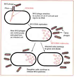
m13_bacteriophageDiagram

The M13 infection cycle, the replication process

  1. In this cycle the DNA is put into the bacteria through the F-pilus.
  2. Once inside the cell the single stranded molecule acts as the template for the synthesis of a complementary strand, resulting in normal double stranded DNA.
  3. This molecule is not inserted in to bacterial genome but instead replicates until over 1000 copies are present in the cell.
  4. When the bacterium divides, each daughter receives copies of the phage genome. This continues to replicate thereby maintaining its overall numbers per cell.
  5. The new phage particles are continuously assembled and released.

m13 life clcleDiagrammatic representation

For further information about M13 bacteriophage click HERE

Plasmids: lecture notes on 17th march

Plasmids

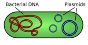
It is an extra chromosomal DNA molecule separate from the chromosomal DNA which is able to replicate independently of the chromosomal DNA.

  • They vary from 1kb to 1000kb
  • The term plasmid was first introduced by the American molecular biologist Joshua Lederberg in 1952

plasmid

  • The plasmid doesnot contribute to the genome of the bacterial cell it is present in but often translates proteins of significance. For example
    • It contains the antibiotic resistance gene that helps the bacteria survive from that specific antibiotic.
    • It can provide the bacteria with an ability to fix elemental nitrogen or to degrade calcitrant organic compounds which provide an advantage under conditions of nutrient deprivation.

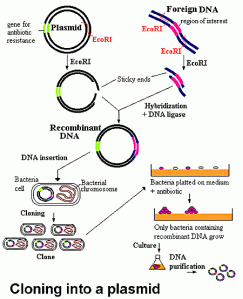
    A small diagram representing the function of the plasmid in gene cloning

gene cloning 2

http://bioweb.wku.edu/courses/biol350/CloningVectEuk9/Review.html

For a better understanding of plasmids, integrative replication and about Ag Tumefaciens.

Replication of plasmid

Non Integrative Replication

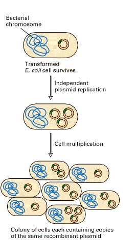
In this type of replication the plasmid DNA once introduced into the cell grows as per the copy number and the multiplication of the cells

non integrative plasmid

Integrative Plasmid

Episome

Under certain conditions some plasmids may integrate into the bacterial chromosome. They are called episome or integrative plasmids. At this stage they replicate along with the bacterial chromosome.

The plasmids in this way are classified into 2 types

Relaxed plasmids

They are the ones which are normally maintained at multiple copies per cell.

Stringent plasmids

They are the ones which have a limited number of copies per cell.

In this case of plasmid replication, the plasmid DNA is integrated in the bacterial chromosome and grows along with the cell. It uses the bacterial machinery for division A good example of this type of replication is Ti Plasmid, often used in agricultural genetic engineering experiments. It completely uses the cell genetic mechanism to grow.

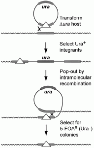
In this case integration and excision of a plasmid is given. Please refer the animation below for better understanding.

integrative plasmid

This animation link given below will help to gain a better understanding of the integration and excision of the vector gene from the chromosomal DNA

http://www.sutree.com/LoadVideo.aspx?s=5073&url=http%3a%2f%2fhighered.mcgraw-hill.com

Types of plasmid

The plasmids are classified as per the conjugation properties as in it was done warlier, but plasmids are now classified using functions

  1. Fertility plasmid:

That contains the tra genes required for conjugation else known as F plasmids. For example F plasmids of E coli

  1. Resistance Plasmid or R plasmids

Carry geens of resistance to one or more antibacterial agents such as ampicilin, mercury etc

  1. Pseudomonas Plasmid:

Such types of plasmids that help in treatment of bacterial infection

  1. Col plasmid:

It contains genes for production of bacteriocins, proteins that kill other bacteria example col E1

  1. Degradative plasmid:

It produces proteins for degradation of unusual substance like toluene and salicylic acid. Example TOI of pseudomonas

  1. Virulent plasmids:

Which in turn convert bacteria into a pathogen. For example, the Ti plasmid of agrobacterium tumefaciens induces crown gal disease on dicot of the palnts

  1. Yeast plasmids:

They are normally 2 micrometer circular plasmids present in yeast cells and other eukaryotic cells

Incase you need a list of plasmids and their sources click       HERE

For more information on plasmid please click                               HERE

Size of some plasmids as vectors

Plasmid

Size

Mol. Wt in kb

Marker

Organism

puc 8

2.1

2.7

Apr

Escherichia coli K12 JM83

col E1

6.4

6.6

E1 imm

Escherichia coli JC411

rP4

54.0

56.4

Apr, Kmr, Tcr

Escherichia coli K12 J53

ToL

117.0

78.0

pseudomonas

pTiach 5

213.0

142.0

Ag. Tumefaciens